In this blog, Lead Scientist Dr Phil Auckland explores how complimentary high-throughput and high-resolution analyses can reveal the mechanisms behind endosomal escape to maximise the cytosolic delivery of RNA therapeutics.
August 2026
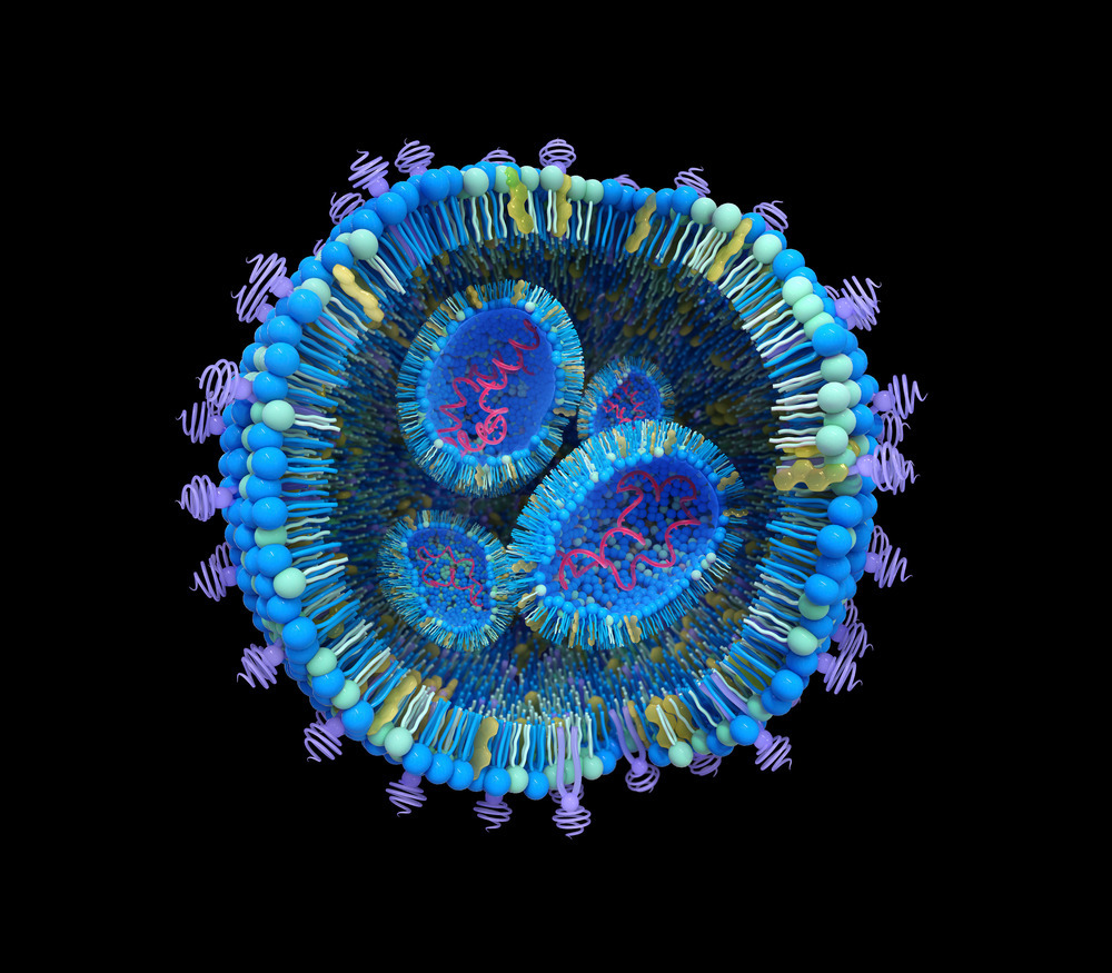
The utility of complex therapeutics directed at intracellular targets is limited because these bulky macromolecules cannot freely translocate the plasma membrane. To overcome this, drugs are often encapsulated within a vehicle that binds cell surface receptors, piggybacks endocytosis for internalisation, then facilitates API escape from the endosome to its cytosolic site-of-action. Many RNA therapeutics are restricted by this permeability barrier, and as a result, are formulated into lipid nanoparticles (LNPs) for intracellular delivery.
Central to LNP functionality are ionisable lipids with a pKa that allows protonation in acidic environments while being neutral at physiological pH. During formulation, these lipids are protonated in acidic buffer to enable electrostatic complexation with the negatively charged RNA payload. Once formulated LNPs are stored and administered at neutral pH where the ionisable lipid is deprotonated. Cytosolic RNA delivery is dependent on this process essentially operating in reverse once LNPs are internalised into endocytic vesicles. As endosomes acidify during maturation from an early to late state (Figure 1a), protonation of the ionisable lipid facilitates its interaction with anionic lipids on the luminal side of the endosomal membrane. This interaction drives the formation of a nonbilayer structure that damages the membrane and allows for RNA escape into the cytosol (henceforth referred to as endosome rupture). Despite being conceptually straightforward this process is highly inefficient, with escape rates estimated at 2-7% using complimentary electron microscopy and live-cell fluorescence approaches. RNA payloads that remain trapped within endosomes are functionally inert, either being returned to the extracellular space by recycling endosomes or destroyed when late endosomes fuse with lysosomes.
Since nearly all drug molecules remain trapped within endosomes, RNA therapeutics are dosed in theoretically excessive quantities, increasing the cost-of-goods and likelihood of off-target or toxic effects. As a result, improving RNA delivery from endosomes, even by single-digit percentage points, is highly attractive to drug developers and underscores the need for empirical strategies to assess cytosolic delivery. At Medicines Discovery Catapult, we approach this in three ways, (1) characterising the dynamics of endosomal rupture, (2) linking rupture dynamics to nanoparticle internalisation and payload expression, and (3), identifying and targeting the specific escape compartment.
To quantitatively assess endosomal rupture and link this to nanoparticle internalisation and payload expression, we established a medium-throughput live-cell imaging assay. Here, LNPs encapsulating mGreenLantern mRNA (mGL, a GFP variant) were labelled after formulation with the RNA dye SYTO60 and transfected into HeLa cells stably expressing Galectin-9-mCherry (Gal9-mCherry), which is specifically recruited to the luminal face of the limiting membrane at rupturing endosomes. This quantitative time-lapse assay enables internalisation to be visualised by SYTO60 translocation into cells, rupture to be visualised by the formation of Gal9-mCherry puncta, and payload expression to be detected by cytosolic mGL fluorescence (Figure 1c).
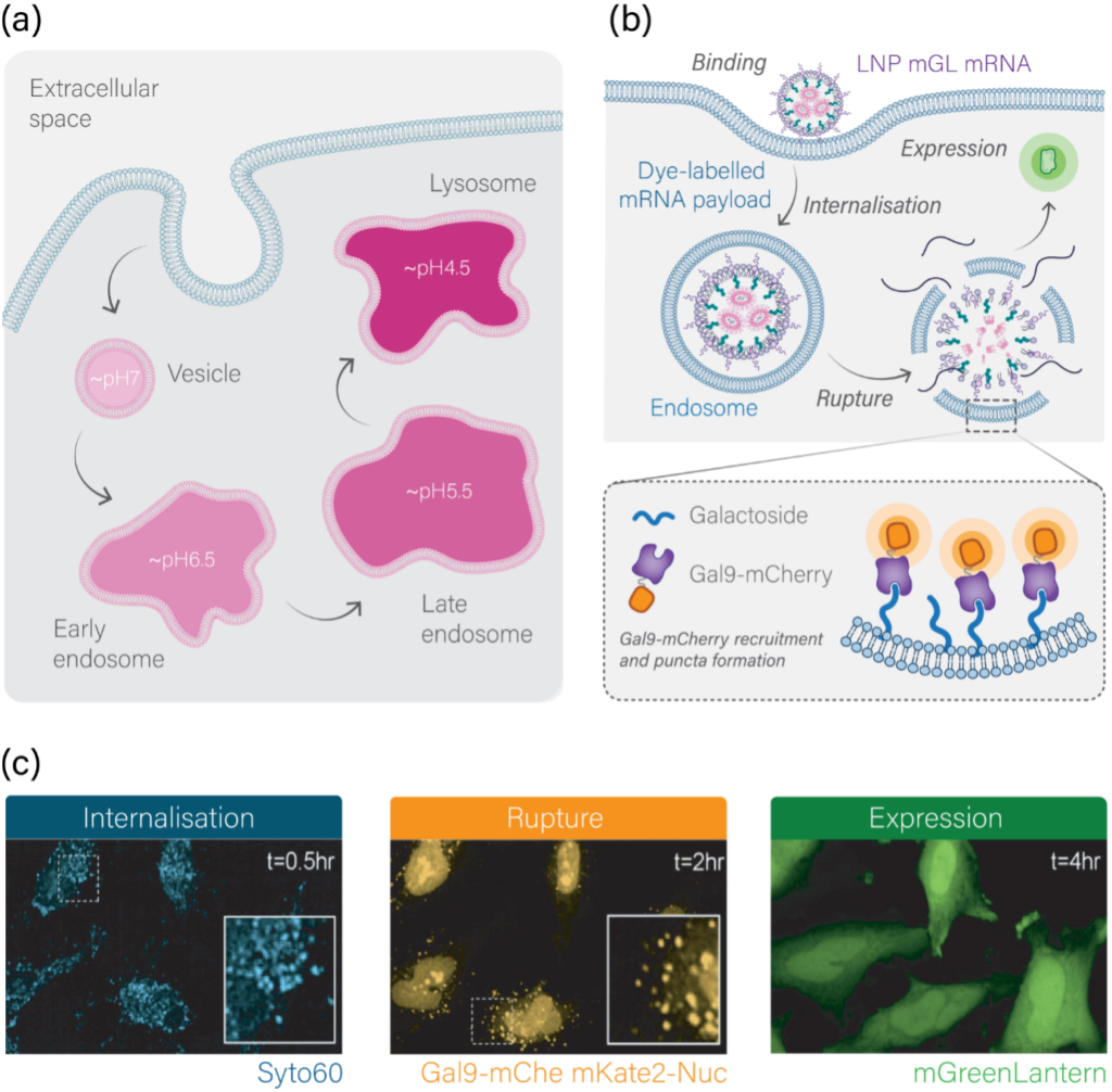
Figure 1: Endosome acidification & the internalisation-rupture-expression assay. (a) Schematic depicting pH of endosomal subcompartments. (b) Schematic showing the internalisation-rupture-expression assay (see text for details). (c) Movies stills of HeLa cells expressing Gal9-mCherry and nuclear-localised mKate2 transfected with LNPs encapsulating mGL mRNA labelled with SYTO60. Snapshots show internalisation (intracellular SYTO60 signal), rupture (Gal9-mCherry puncta), and payload expression (cytosolic mGL protein).
To explore how LNP composition directs internalisation-rupture-expression behaviour, 39 LNPs encapsulating mGL mRNA were formulated at the Centre for Process Innovation (CPI) and screened using the live-cell imaging assay. Our data revealed significant variation in the kinetics and magnitude of internalisation-rupture-expression behaviour between formulations (Figure 2a-c). Moreover, plotting the maximal effect for each parameter showed how these processes are not directly related, for example, some formulations that highly internalise fail to effectively rupture endosomes (Figure 2d).
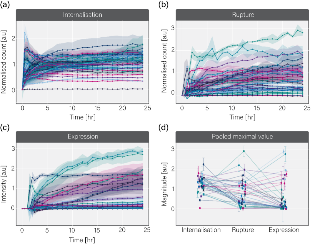
Figure 2: Pooled internalisation-rupture-expression kinetic data for 39 LNP formulations. Traces showing the internalisation (a), endosome rupture (b), and mGL expression kinetics (c) for 39 LNP formulations encapsulating mGL mRNA, labelled with SYTO60 and transfected into HeLa cells expressing Gal9-mCherry. (d) The maximum internalisation, rupture and expression value for each tested LNP formulation. Lines link the same formulation across parameters. Error bars/shading = +/-SD.
Because payload expression is a function of both nanoparticle internalisation and endosomal rupture we sought to understand the predictive value of each metric. Plotting maximal internalisation and rupture against mGL expression revealed meagre positive correlations (R2 of 0.1 and 0.23, respectively, Figure 3a,b). In contrast, combining internalisation and rupture into a single index metric to approximate payload delivery significantly increased predictive value and identified three distinct formulation classes (Figure 3c). These are: (1) formulations with poor delivery and poor expression, (2) formulations with high delivery and high expression, and (3), formulations with high delivery and poor expression. Classes (1) and (2) represent a formulation space where this assay has high utility, since effective delivery is directly associated with payload expression and vice versa (Figure 3c). Conversely, class (3) identifies atypical formulations, which appear to internalise into cells and rupture endosomes but fail to express their payload. We note this observation cannot be explained by physicochemical defects or RNA integrity.
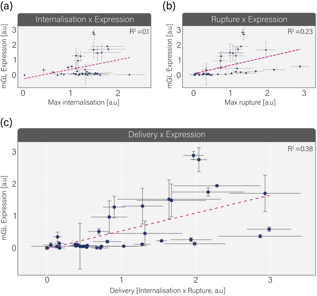
Figure 3: Combing internalisation and rupture into a predictive metric termed delivery. (a) Scatter plot of maximal internalisation against maximum payload expression. (b) Scatter plot of maximal rupture against maximum payload expression. (c) Scatter plot of delivery (normalised internalisation x rupture) plotted against maximum payload expression. Error bars = +/-SD. Dotted line indicates the linear regression fit used to calculate R2.
Comparing delivery against expression suggested that some apparently high performing formulations (high delivery) fail to express their mRNA payload. A possible explanation for this is the spatial resolution at which the assay is conducted. In medium-throughout format rupture is analysed on a whole cell basis, with the number of rupture events quantified per cell over time. However, rupture is a subcellular event directed by the asynchronous timing of endosome formation (Figure 4a), therefore, to shed light on the high delivery-low expression paradox, we visualised endosome rupture and payload dynamics with single-endosome resolution. To this end, HeLa cells stably expressing Gal9-GFP were transfected with nanoparticles encapsulating Cy3-labelled siRNA and imaged every 3min for 12hr using high-resolution confocal microscopy. We then quantified the intensity of endosomal siRNA-Cy3 over time as an endosome underwent rupture and recruited Gal9-GFP. This revealed two rupture subtypes, one where the siRNA-Cy3 signal was lost as the endosome underwent rupture and the payload escaped, and another where siRNA-Cy3 persisted in the endosome despite rupture and Gal9-GFP recruitment (Figure 4b,c). These opposing fates were dynamically separable; escape was associated with faster rupture following internalisation and a higher Gal9-GFP:siRNA-Cy3 ratio at rupture onset (Figure 4d,e). Altogether, these data demonstrate that rupturing endosomes do not always release payload molecules, and that payload escape is associated with early rupture that exposes a large proportion of the endosomal leaflet to the cytosol.
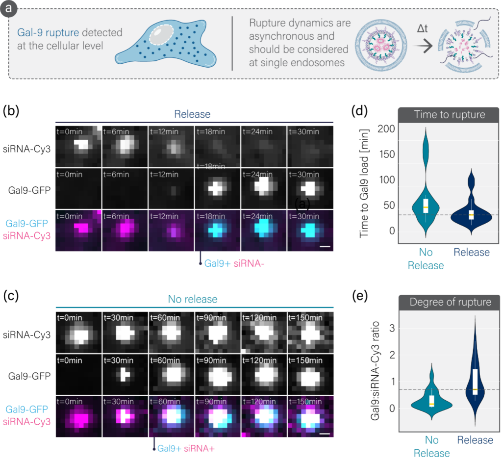
Figure 4: Studying endosomal rupture at the level of individual endosomes. (a) Comparison of assay approaches, where medium throughput considers rupture across the entire cell while high-resolution analysis considers rupture and payload fate at single endosomes. (b) Movie stills of endosomal rupture (Gal9-GFP recruitment) leading to release of the siRNA-Cy3 payload into the cytosol. (c) Movie stills of siRNA-Cy3 payload molecules remaining trapped within an endosome despite it undergoing rupture and recruiting Gal9-GFP. (d) The time between internalisation and rupture for endosomes that release their payload and those that do not. (e) The Gal9-GFP:siRNA-Cy3 ratio at rupture onset for endosomes that release their payload and those that do not. We suggest this reflects the degree of membrane disruption for a given amount of payload.
A final question pertains to the identity of endosomes that release payload molecules upon rupture. To explore this, we used CRISPR-Cas9 genome engineering to knockin mStayGold (mSG) at the endogenous loci of Rab7a, a marker of late endosomes, in cells expressing Gal9-mCherry. When transfected with nanoparticles containing siRNA-647, this enables the rupture-escape relationship to be studied in the context of endosomal identity (Figure 5a). By using mSG-Rab7a as a molecular clock, we quantified payload escape at endosomes that ruptured in an early state, before becoming a Rab7a positive compartment, or after maturation into a late Rab7a-bound vesicle. We found that approximately 70% of payload molecules escaped from rupturing early endosomes, while only 30% of payload molecules escaped from rupturing late endosomes (Figure 5b-d). This demonstrates the existence of a delivery window where payload escape-compliant rupture occurs, which is associated with early endosomal vesicles and requires high-resolution analysis for detection (Figure 5e).
So, how to study the endosomal escape of RNA therapeutics? Developers must first understand how their formulation space impacts the kinetics of internalisation-rupture-expression. This will identify the parameters that direct payload expression and, importantly, provide granular experimental insight into outliers. From here, the focus should shift onto payload tracking in the context single endosomes to support the iterative optimisation of vehicle technology to maximise productive rupture. The latter is increasingly important given recent findings of rupture-induced inflammation, which attenuates payload efficacy and provides a strong argument for avoiding therapeutically inactive rupture events.
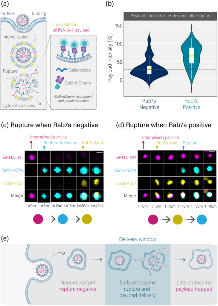
Figure 5: Identification of the escape window. (a) Schematic depicting the assay used to understand how endosome identify directs rupture productivity. (b) The percentage of payload molecules remaining in the respective endosome types following rupture. (c) Movie stills of an siRNA-647 containing endosome undergoing rupture before mSG-Rab7a loading. (d) Movie stills of an siRNA-647 containing endosome undergoing rupture after becoming a late mSG-Rab7a positive compartment. (e) Schematic depicting the delivery window where escape-compliant rupture occurs.
MDC’s preclinical cascade combines high-throughput formulation screening, mechanistic intracellular delivery characterisation, and in vivo biodistribution to build experimental confidence at every stage.
Speak to our team to find out how we can accelerate your path to the clinic.


Xenium at MDC Case Study: Hypoxia in Head and Neck Cancer
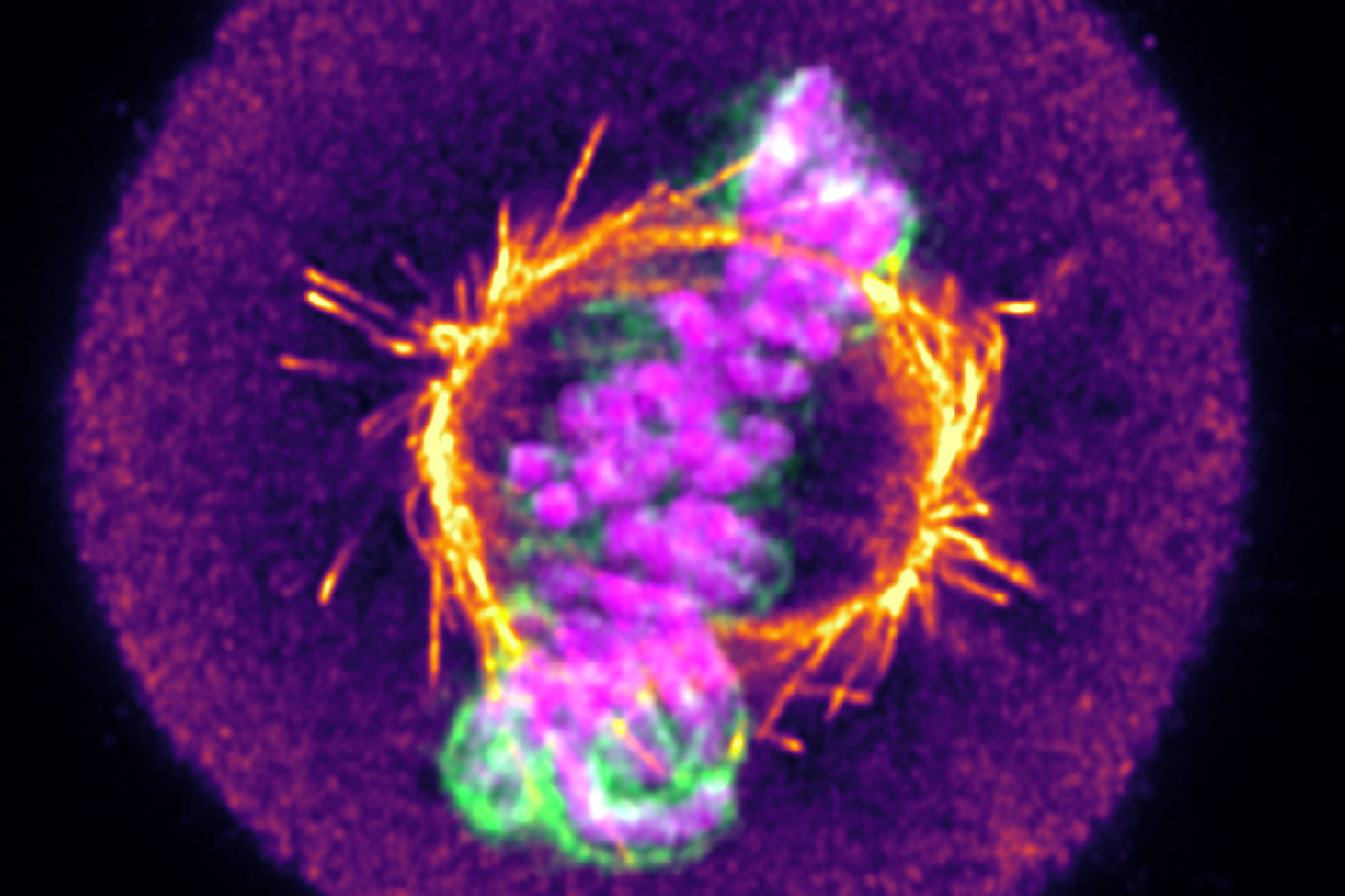
From mitochondrial networks that silently rebalance fusion and fission, to chromosomes that only reveal their fate in the minutes after division, live-cell imaging uncovers dynamics that static snapshots miss entirely. We explore three examples, including how tracking RNA therapeutic vehicles as they transit and escape endosomal compartments in real time resolves ambiguity that even super-resolution imaging of fixed cells can’t overcome.
By looking beyond the diffraction limit, we have established a method to quantitatively distinguish low-grade breast cancers that appear identical by traditional means. Ultimately, this nanoscale insight could expand access to novel HER2-targeted therapies by reliably identifying low expressing cohorts that would benefit from intervention.