What does hypoxia look like inside a tumour, cell by cell? Our team can map it, single-cell spatial transcriptomics for up to 5,000 targets in standard FFPE tissue.
Below is a head and neck tumour core, part of a larger study with Manchester Cancer Research Centre exploring hypoxia’s effect on the tumour microenvironment, using the 10x Genomics Xenium platform.
July 2026
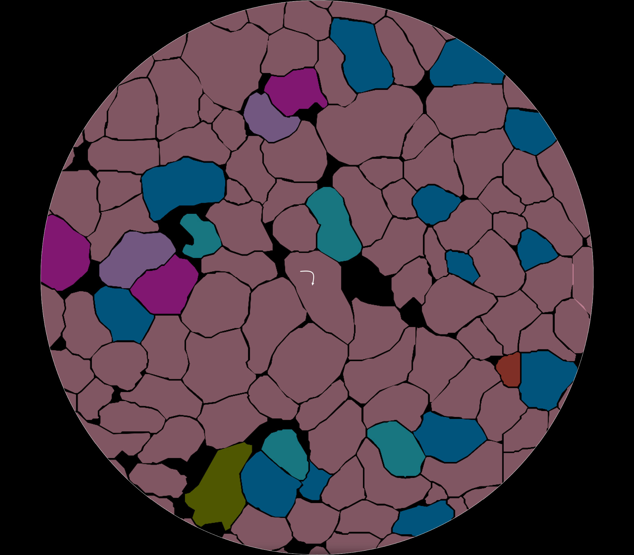
Molecularly profiling up to 5,000 transcripts at single-cell level, in FFPE tissue.
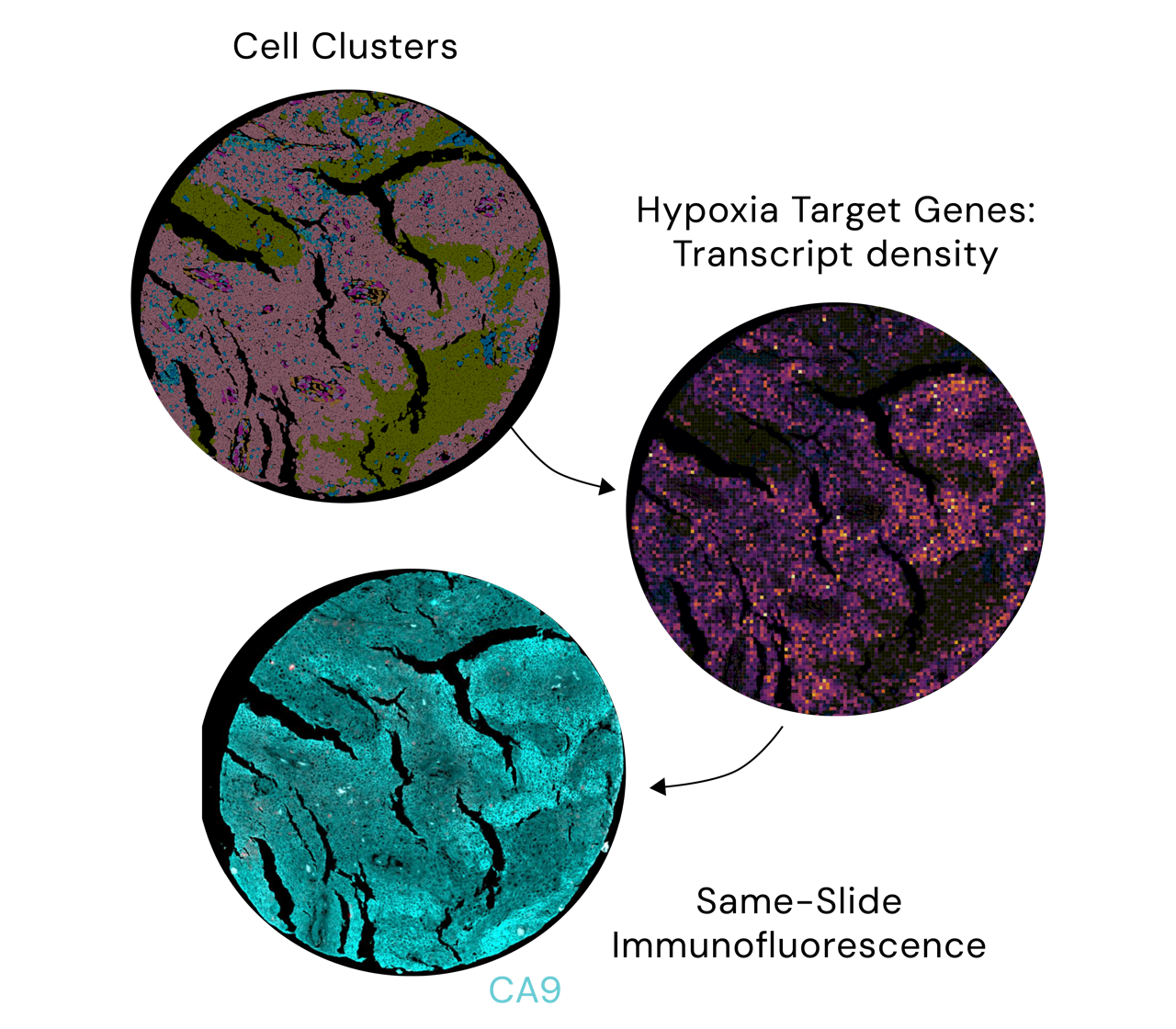
Using the Xenium platform, we mapped the genes driving the hypoxic response at single-cell resolution, then confirmed the presence of hypoxia in those same regions using CA9 immunofluorescence staining, on the very same slide.
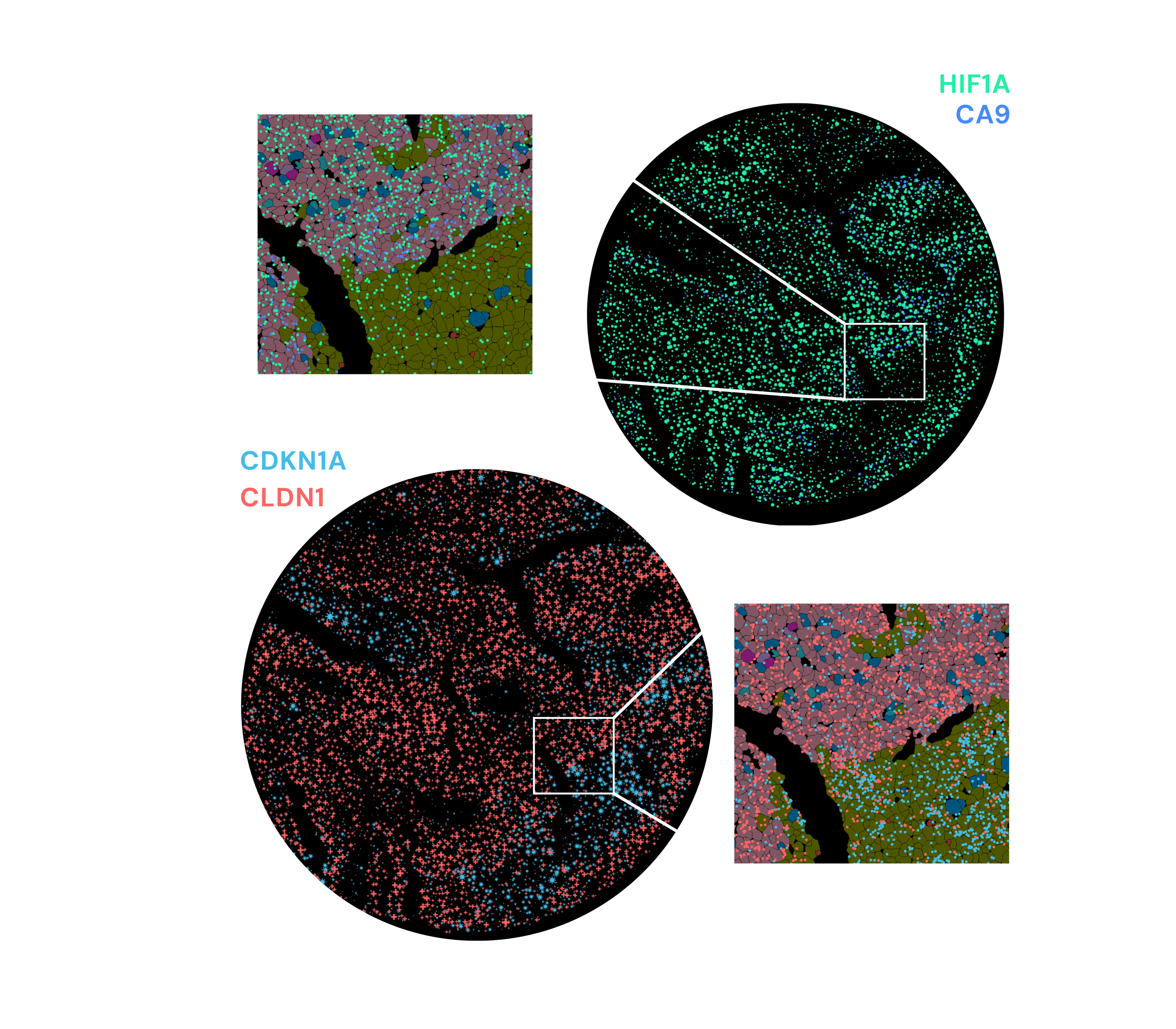
These genes corresponded to different subtypes of tumour cells within the same tumour, mapped to their exact position, cell by cell.
It can also provide insights across immunology, neuroscience, and other oncology programmes, Xenium has joined the ranks of our spatial biology platforms at MDC, alongside GeoMx and CosMx.
If your project could benefit from single-cell spatial resolution, we’ll help you work out whether it’s the right fit, and which of our platforms answers your question best.




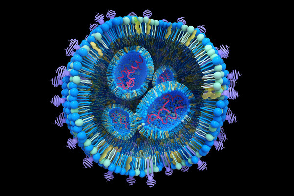
How to study the endosomal escape of RNA therapeutics
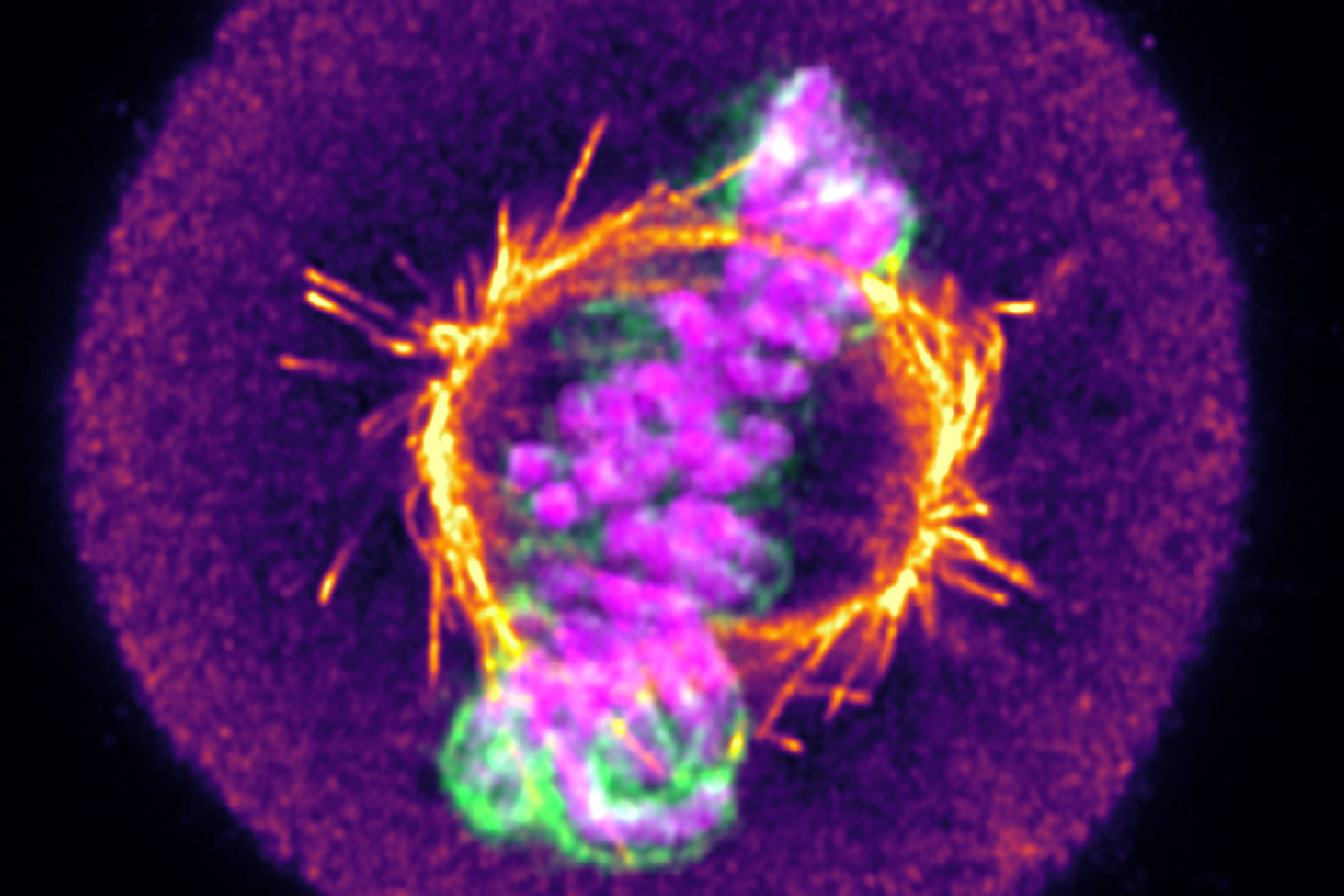
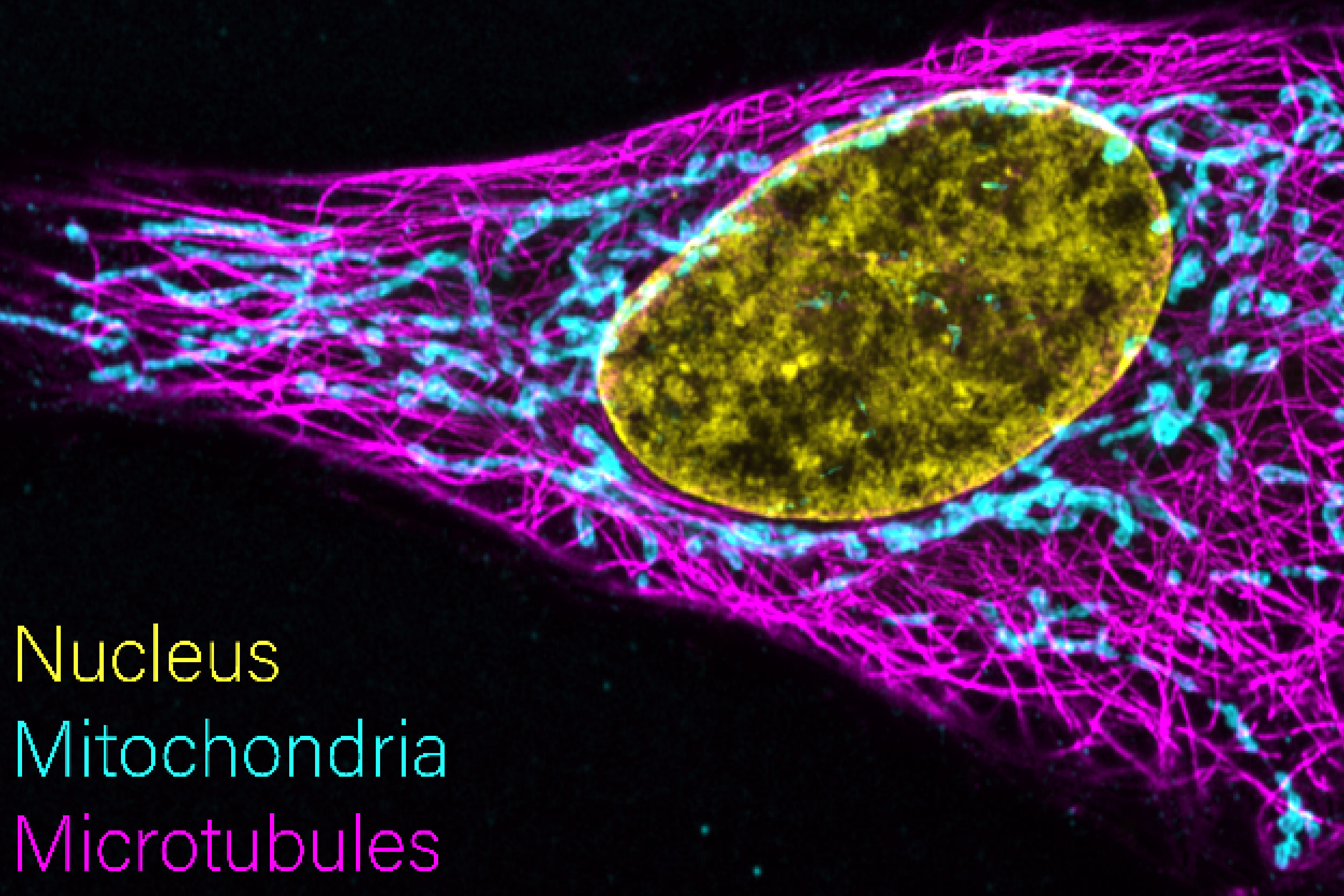
From mitochondrial networks that silently rebalance fusion and fission, to chromosomes that only reveal their fate in the minutes after division, live-cell imaging uncovers dynamics that static snapshots miss entirely. We explore three examples, including how tracking RNA therapeutic vehicles as they transit and escape endosomal compartments in real time resolves ambiguity that even super-resolution imaging of fixed cells can’t overcome.
By looking beyond the diffraction limit, we have established a method to quantitatively distinguish low-grade breast cancers that appear identical by traditional means. Ultimately, this nanoscale insight could expand access to novel HER2-targeted therapies by reliably identifying low expressing cohorts that would benefit from intervention.