February 2020
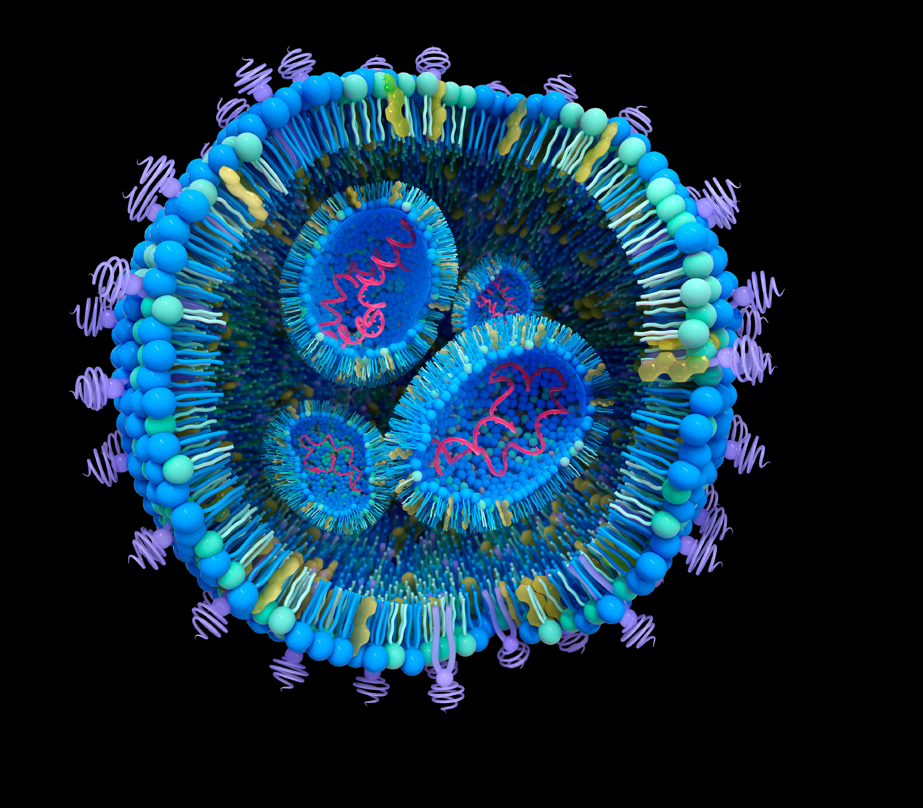
One of the most important challenges in drug development is efficient delivery of the drug molecule to the appropriate tissue to drive therapeutic benefit, while minimising side effects. For small-molecule drugs, a suboptimal ‘therapeutic index’ between the doses that deliver desired and undesired effects can limit effectiveness or even halt progression through clinical trials. For non-small-molecule drugs, such as antibody-drug conjugates and oligonucleotides, their high molecular weight and other properties can limit their distribution throughout the body, thereby preventing the drug from reaching its target and exerting a therapeutic effect.
Imagine an anti-cancer drug designed to kill a tumour. When given to the patient, this drug molecule needs to move through the circulatory system, navigate the extracellular environment, penetrate the tumour tissue before it gets removed and metabolised and finally enter the cell and bind to its target. All while causing minimum effect on normal cells.
Nanotechnology-based drug delivery systems hold tremendous potential to overcome these challenges. Designed to carry the drug(s) or genes specifically to the target cells or tissues, the systems release it at the desired concentrations, decreasing undesired toxicity effects. This allows for rapid evaluation of treatment efficacy in the early stages.
Over the last decade, dozens of nanomedicines have received clinical approval, with many more in preclinical development or in clinical trials.
For nanomedicines to be successful in the clinic, an in-depth mechanistic understanding of these delivery systems is crucial. There are several critical questions that need answers:
Answering these questions will help to develop an effective drug delivery system. Once the delivery mechanism is understood, the properties of the system (size, surface charge, shape, hydrophobicity, surface chemistry) can be tailored to evade biological barriers and achieve optimal cellular uptake across different organ systems to treat various diseases.
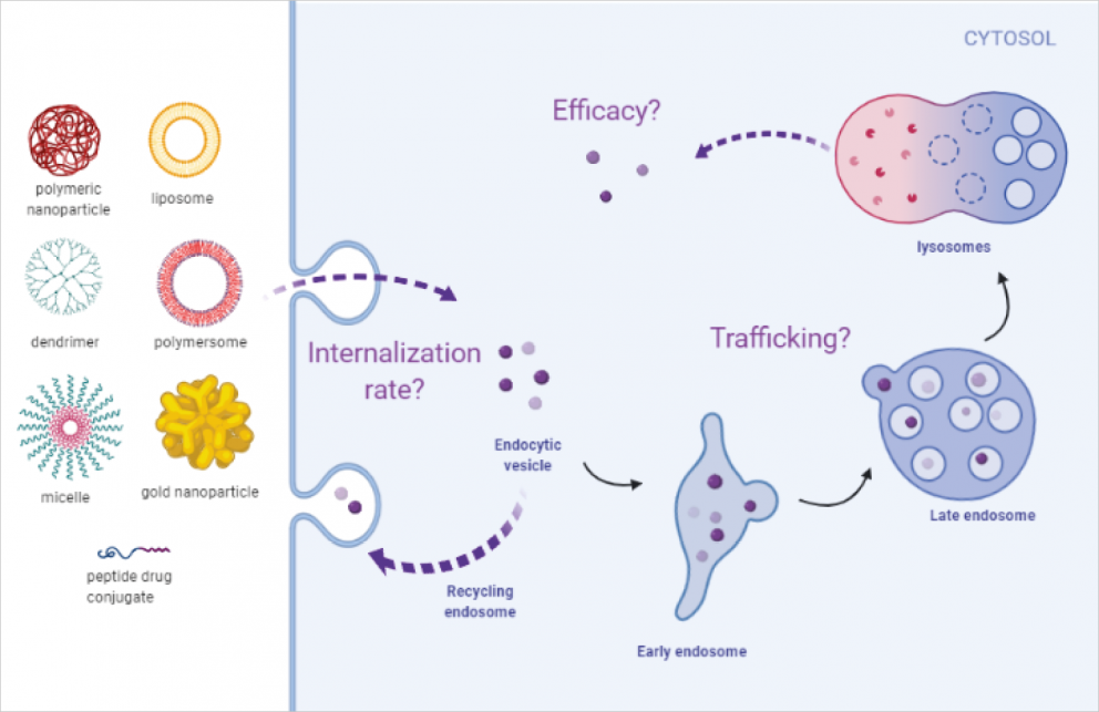
Cellular entry and trafficking of nanomedicines
At Medicines Discovery Catapult, we have developed a microscopy-based platform to study the cellular entry and trafficking of nanomedicines. This image-based platform provides a sensitive and robust method for evaluating nanomedicines in cellular systems and enables further improvement of drug delivery systems.

Dr Duygu Yilmaz, PhD, is a Senior Scientist in Target Engagement & Validation at Medicines Discovery Catapult, with over 5 years’ experience in drug research and development industry.
During her PhD, Duygu worked on functionalised liposomal drug delivery systems, which would release the drug payload in a highly sensitive manner in response to stimulus in the environment. She has experience in various other drug delivery systems including polymeric nanoparticles and cell penetrating peptides. At MDC, she is developing microscopy-based methods to study intracellular entry and trafficking of nanoparticles.
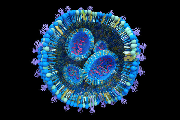
How to study the endosomal escape of RNA therapeutics
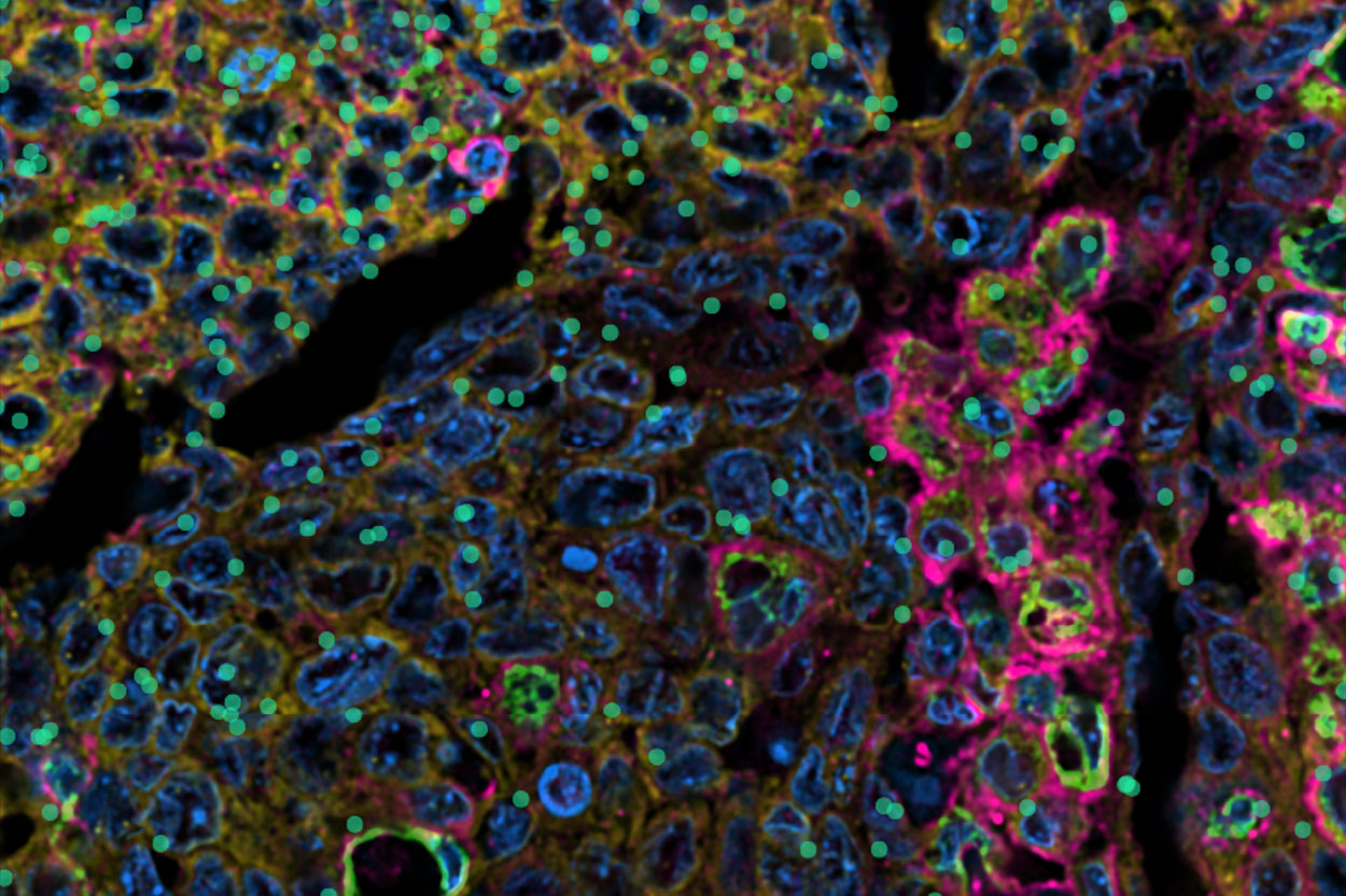
Xenium at MDC Case Study: Hypoxia in Head and Neck Cancer
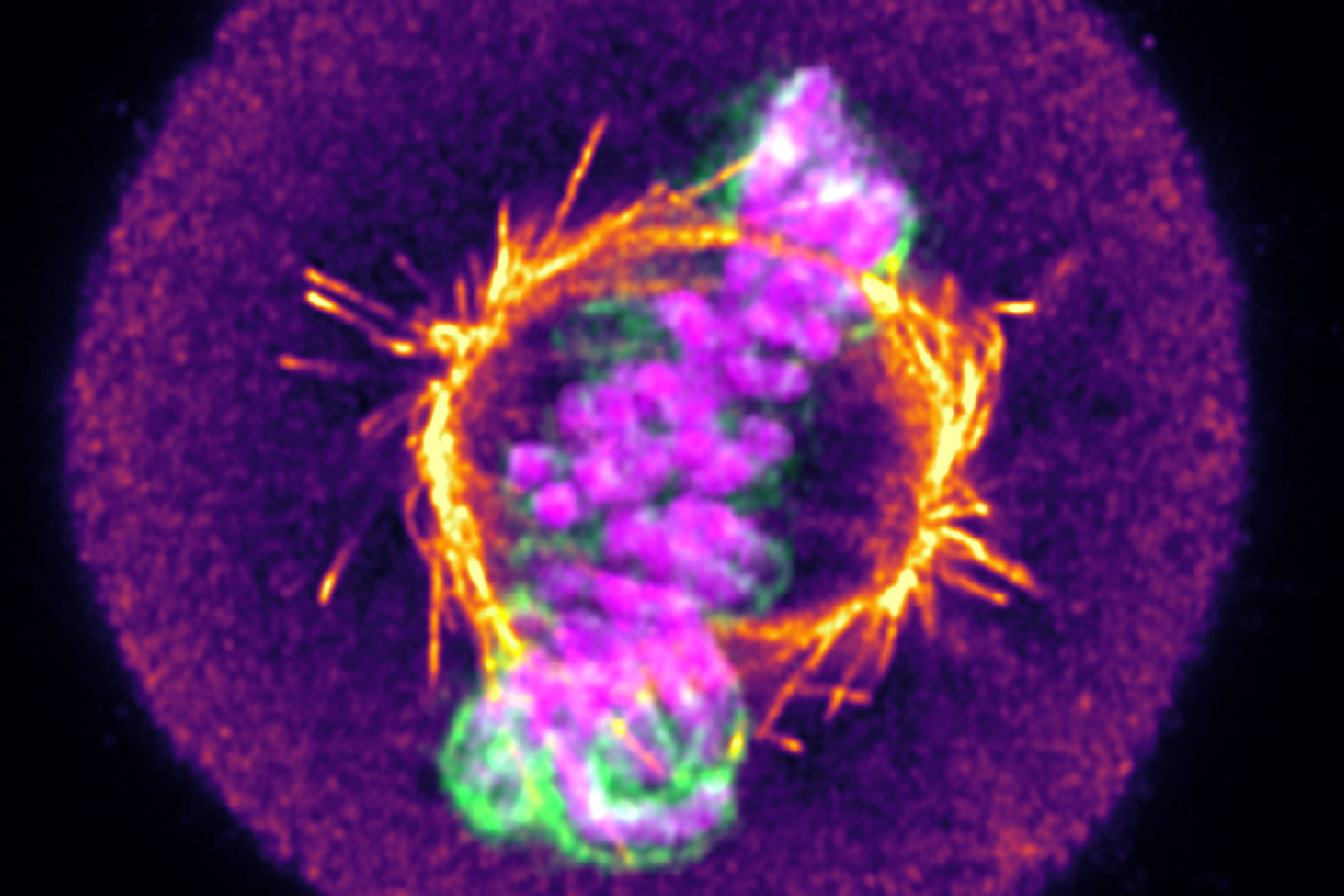
From mitochondrial networks that silently rebalance fusion and fission, to chromosomes that only reveal their fate in the minutes after division, live-cell imaging uncovers dynamics that static snapshots miss entirely. We explore three examples, including how tracking RNA therapeutic vehicles as they transit and escape endosomal compartments in real time resolves ambiguity that even super-resolution imaging of fixed cells can’t overcome.