June 2026
The RNA therapeutic market has surged since 2021, driven primarily by the increase in mRNA and siRNA therapies in development (Figure 1a). These drugs target prevalent and diverse pathologies, from gene therapies for rare conditions to anticancer agents and cardiovascular drugs (Figure 1b). Such widespread application has necessitated the development of delivery technologies capable of protecting delicate RNA payloads and targeting them to disease-associated cells in a programmable and precise manner. As such, tools capable of quantitatively evaluating payload effect with cell type specificity in tissues isolated from model systems have become evermore valuable. These empower innovators to maximise on-target efficacy and preclinical translatability by providing a granular understanding of vehicle-payload biodistribution and target modulation.
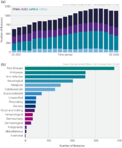
Figure 1. The RNA therapeutic market from modality (a) and disease area (b) perspectives.
LNPs are recognised as a leading delivery platform following clinical success in vaccine and therapeutic applications. LNPs are made by combining neutral and sterol lipids with an ionisable lipid that encapsulates the RNA payload and a PEG lipid that reduces aggregation and extends circulation time (Figure 2). Medicines Discovery Catapult (MDC), as part of the Intracellular Drug Delivery Centre (IDDC), has built a preclinical platform to evaluate LNPs against two major challenges in the field, (1) limited cytosolic payload delivery, and (2) programmable tropism in vivo. To help address the latter, MDC has established a deep learning-based immunofluorescence workflow to quantify mRNA payload expression with single-cell resolution ex vivo. Below, we present a case study of this workflow using intravenously administered LNPs encapsulating mGreenLantern (mGL, a GFP variant) mRNA, which differentially target four major liver cell types (Figure 2).
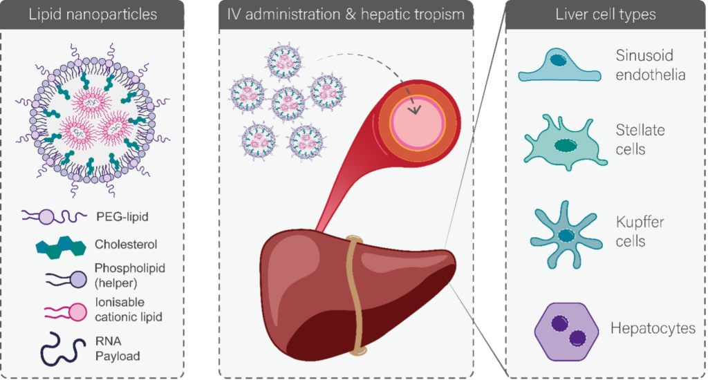
Figure 2: The composition and fate of liver-tropic LNPs. Left Lipid nanoparticles are made by combining structural phospholipid and sterol components with an ionisable lipid that encapsulates the RNA payload and a PEG lipid that reduces aggregation and extends circulation time. Middle & right When administered intravenously, many lipid nanoparticles display strong hepatic tropism through binding of apolipoprotein E, ultimately delivering their payload to one of the four constituent liver cell types.
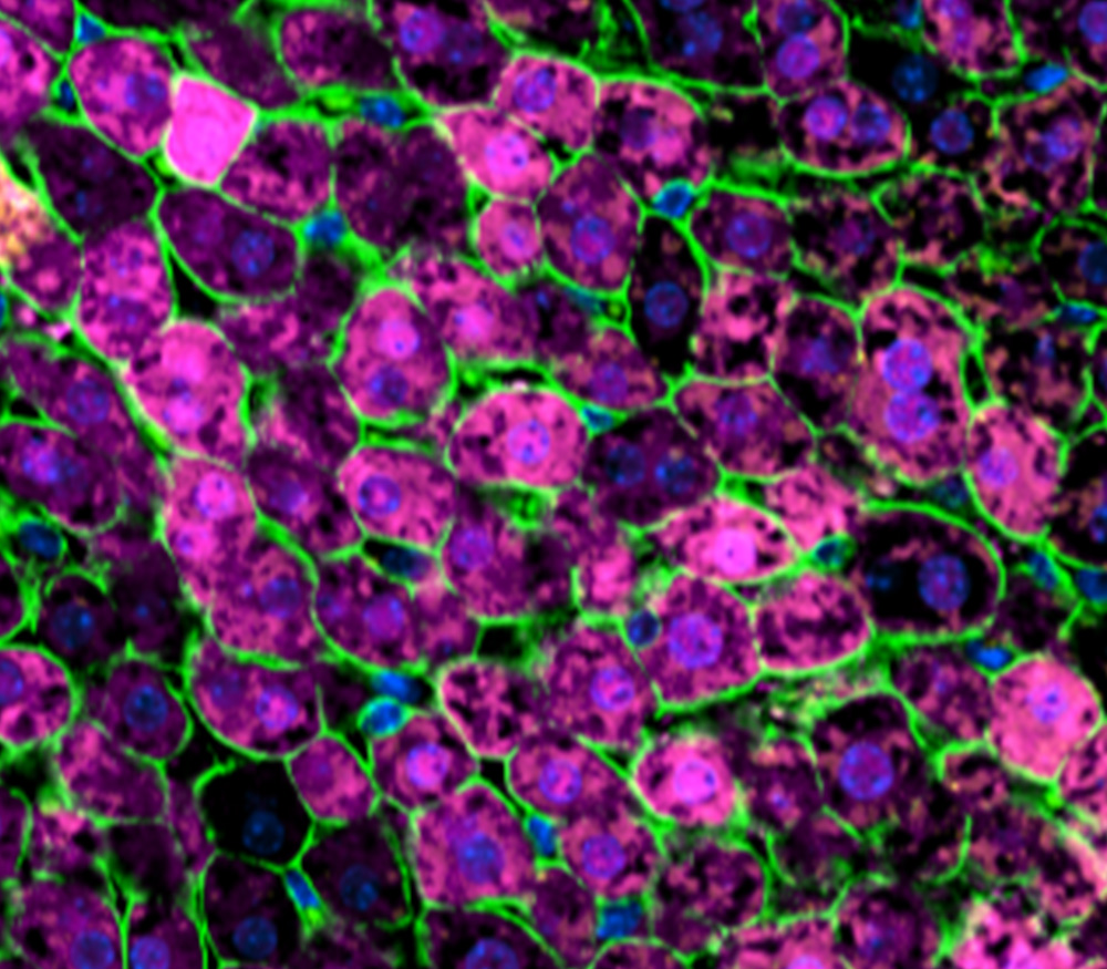
To evaluate mGL expression in distinct liver cell populations ex vivo we built an immunofluorescence workflow with automated sample preparation, imaging, and analysis (Figure 3a-d). Here, FFPE liver sections were prepared from mice treated with a clinical LNP encapsulating mGL mRNA and stained with DAPI and antibodies against mGL, N-cadherin, and a cell type marker. A deep-learning model was trained in Visiopharm to segment the morphologically diverse cell types based on marker staining, from which a mask was created to calculate the mGL positivity rate for the respective cell population (Figure 3c,d). The approach was validated using a panel of clinical LNP formulations encapsulating mGL mRNA dosed at three time points (Figure 4). This demonstrated that mGL mRNA expression is a function of formulation, time, and cell type, and validated the assay for precise quantification of payload expression with cellular resolution (Figure 4).
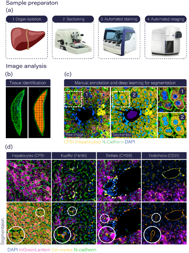
Figure 3: A workflow for automated preparation, imaging, and analysis of cellular tropism in murine liver sections. (a) Sample preparation. (b) Automated tissue region detection with edge exclusion. (c) Training a deep learning model for segmentation of hepatocytes based on CPS1 staining. Zoom boxes depict show the raw immunofluorescence image and the manual segmentation of that region used for training, respectively. (d) Example images showing immunofluorescence staining for all four main liver cells types (top row) and the deep-learning based segmentation of that cell type using the marker staining. Zooms show individual segmented cells.
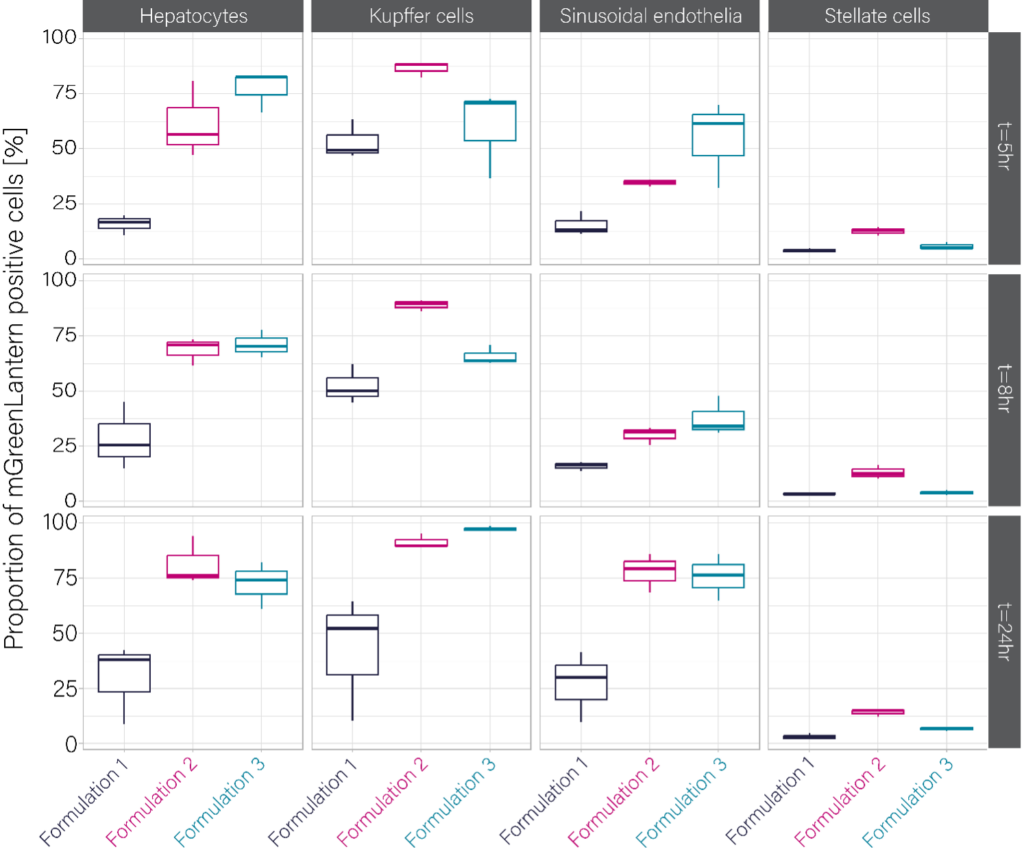
Figure 4: Evaluation of clinical LNP formulations encapsulating mGL mRNA across time points and cell types.
Our analysis has shown how key factors, such as formulation and cell type, control general expression trends. For example, hepatocytes and Kupffer cells are more readily transfected than hepatic stellate cells, and formulations 2 and 3 outperform formulation 1 in all cell types. Nevertheless, given the scope of target cells, developers must look beyond trends and instead optimise formulation to direct transfection toward a single cell type. We therefore sought to demonstrate our workflow was suitable for this purpose using a well-established example; that PEG can control cellular LNP tropism between hepatocytes and Kupffer cells. Mice were intravenously administered LNP -PEG or LNP +PEG particles encapsulating mGL mRNA and liver sections prepared at 24 hr. mGL positivity was quantified in hepatocytes and Kupffer cells as above, revealing a striking reciprocal relationship where LNP +PEG particles primarily transfected hepatocytes while LNP -PEG particles primarily transfected Kupffer cells (Figure 5).
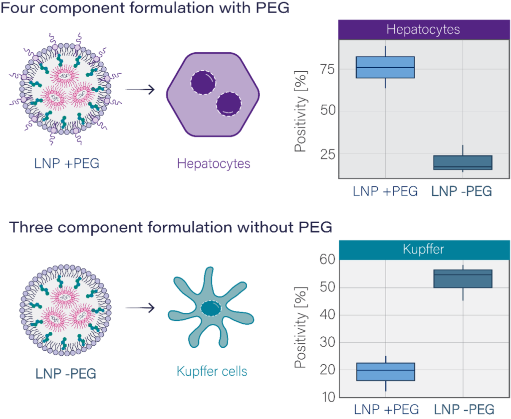
Figure 5: Directing LNP cellular tropism with PEG. PEG-containing LNPs primarily transfect hepatocytes (above) while LNPs formulated without PEG primary transfect Kupffer cells (below).
In summary, we present an immunofluorescence workflow supported by deep-learning image analysis to evaluate cellular tropism in ex vivo tissue. The approach is readily applicable to other tissue types, delivery vehicles, and payloads. By enabling innovators to quantify targeting with such granularity, this method will help optimise delivery vehicles for the precise tropism required to realise the full potential of RNA payloads.
MDC’s preclinical cascade combines high-throughput formulation screening, mechanistic intracellular delivery characterisation, and in vivo biodistribution to build experimental confidence at every stage.
Speak to the team to learn how we can accelerate your path to the clinic.


How to study the endosomal escape of RNA therapeutics
Xenium at MDC Case Study: Hypoxia in Head and Neck Cancer
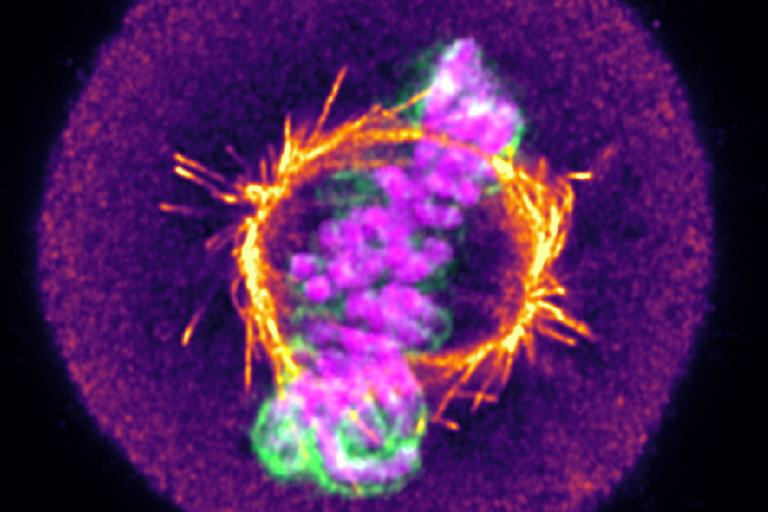
From mitochondrial networks that silently rebalance fusion and fission, to chromosomes that only reveal their fate in the minutes after division, live-cell imaging uncovers dynamics that static snapshots miss entirely. We explore three examples, including how tracking RNA therapeutic vehicles as they transit and escape endosomal compartments in real time resolves ambiguity that even super-resolution imaging of fixed cells can’t overcome.