In vivo imaging to understand the biodistribution of a candidate compound

How imaging technologies can be used to establish the biodistribution and accumulation of the compounds in vivo


Using imaging in drug discovery

Multimodal and multiscale imaging solutions enable quantification and understanding of organ accumulation and biodistribution, target expression and engagement, optimal biological dose, toxicities/drug interactions and preclinical studies to predict what is seen in the clinic.

Overcoming the challenge of quantitatively determining organ accumulation and distribution at specific time points

In vivo and ex vivo preclinical imaging offers a compelling approach and solution to non-invasively characterise and validate compound distribution and accumulation.

Medicines Discovery Catapult (MDC) has imaging expertise in multi parametric imaging modalities:

  • Functional radiological imaging techniques such as positron emission tomography (PET)
  • Computed tomography (CT)
  • High-frequency ultrasound (HFUS)
  • Bioluminescence/near-infrared imaging (NIR)
  • ex vivo imaging techniques:
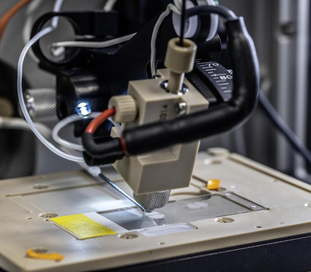
    • Mass spectrometry imaging (DESI and MALDI)
    • Digital spatial profiler (nanostring DSP)
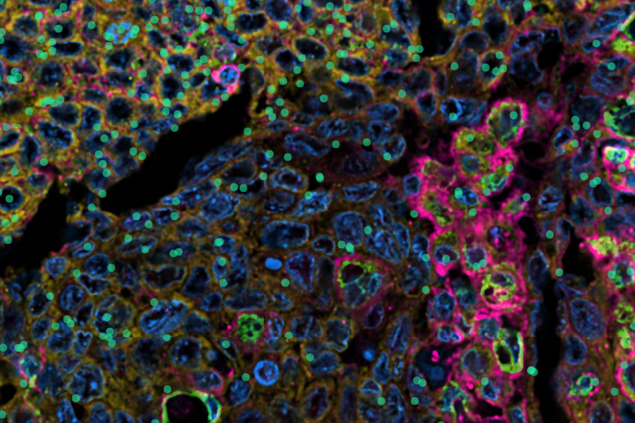
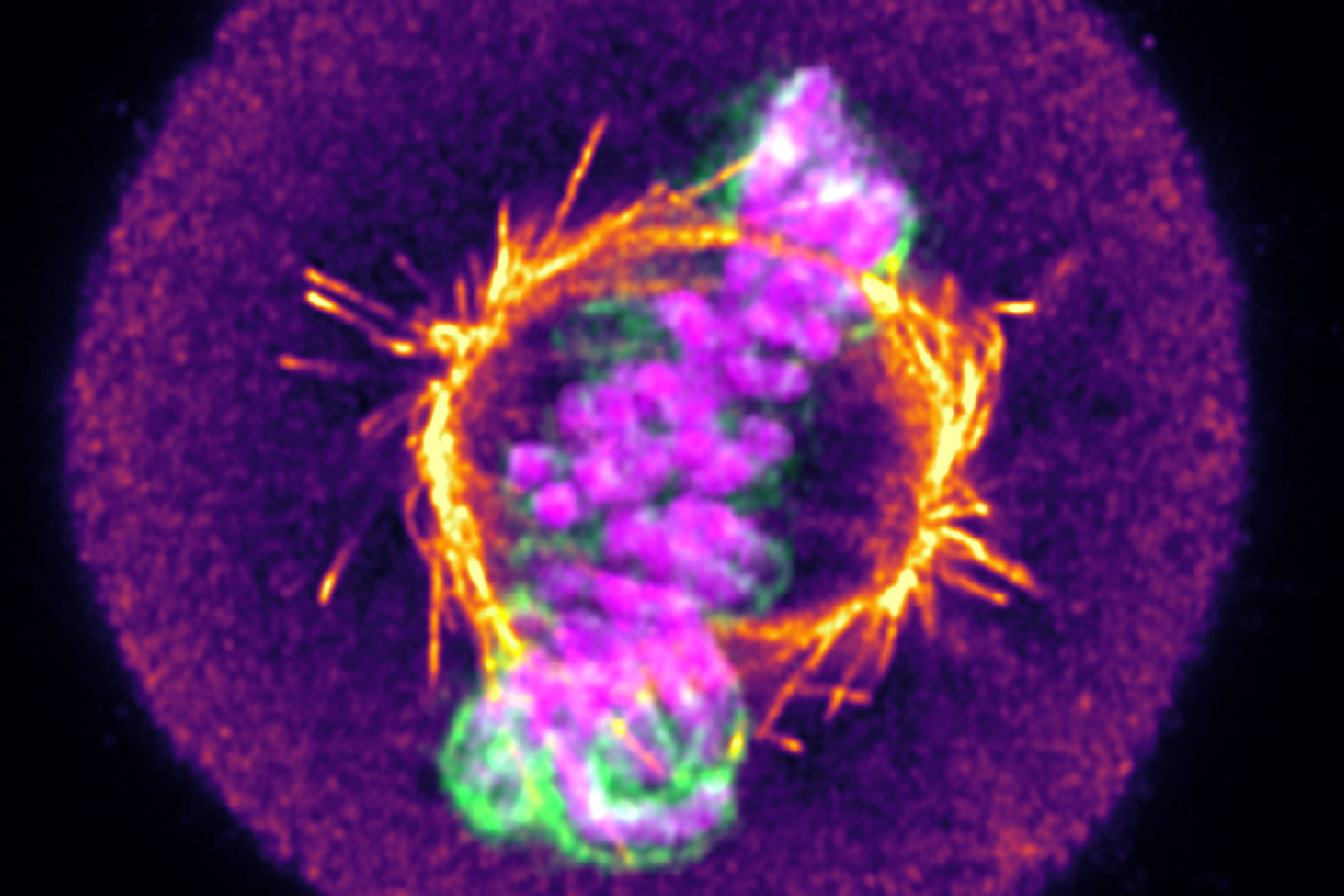
    • Advanced microscopy imaging suite (super resolution/confocal/multiphoton).

How can imaging be used to understand distribution of a therapeutic?

The rate and degree of drug distribution reflects the extent it is present in extravascular tissues and depends on blood flow, capillary permeability, and protein binding.  The administered substance goes through a series of cascades to be effective and imaging can track the progress from the systemic level to the cellular level to understand and characterise it.

The 5 steps are:

  1. Circulation
  2. Accumulation
  3. Penetration
  4. Internalisation
  5. Release

Examples of imaging techniques that can be used include:

  • Fluorescence near infrared imaging device for longitudinal Cy-7 biodistribution. Cy-7 imaging allows deep biological penetration within the body and the ability to image the whole-body distribution with good signal-to-noise. There are many ways to label a molecule with a dye enabling it to be tracked.
  • 89Zr PET can also be used to look at whole body distribution and also off target toxicity and nonspecific binding. 89Zr PET is an ideal tracer due to its half-life, allowing repeated images to longitudinally assess distribution of the compound. This technique can also be used to assess tumour penetration longitudinally.
  • MDC’s collaboration with the University of Leeds and the therapeutic microbubble consortium in cancer nanoparticles uses an enhanced tumour delivery of microbubbles as a drug carrier in combination with a HFUS platform. Using imaging it can characterise the delivery system and demonstrate that accumulation and distribution of the microbubbles are predominantly through the reticulo endothelial system (RES).
  • Three imaging techniques can be used to assess distribution and penetration across the blood-brain barrier (BBB) including PET, near-infrared imaging and bioluminescence probes offer a non-invasive method, and mass spectrometry imaging which has the potential to define drug distribution in very small brain structures with a non-labelled approach.
  • Non-invasive mass spectrometry imaging has broad potential in the drug distribution space. Two main mass spectrometry imaging techniques used include desorption electrospray ionisation (DESI) and matrix assisted laser desorption ionisation (MALDI).

Summary

Imaging at MDC offers huge potential as a platform to non-invasively measure the pharmacokinetics (PK) and characterisation of small molecules, biologics and, non-biological complex medicines to facilitate faster translation into clinical development.

This article is based on Juliana’s talk from the MDC Connects webinar series. Watch the session Juliana took part in –Optimising the Compound:


A photo of Dr Juliana Maynard, de-risking radiopharmaceutical investment using her preclinical imaging expertise at Medicines Discovery Catapult.

Juliana Maynard is Lead Scientist in Pre-clinical Imaging at MDC. She works with a wide range of molecular imaging modalities including PET, SPECT, CT and ultrasound. Previously she was Head of Imaging Services at Alderley Imaging and worked at AstraZeneca for 9 years. Juliana has a PHD in neuroendocrinology from the University of Edinburgh.


Other Blogs

Blogs
05 July 2026
From Photons to Phenotype

By looking beyond the diffraction limit, we have established a method to quantitatively distinguish low-grade breast cancers that appear identical by traditional means. Ultimately, this nanoscale insight could expand access to novel HER2-targeted therapies by reliably identifying low expressing cohorts that would benefit from intervention.


Cellular tropism microscopy liver cell image
Blogs
15 June 2026
Evaluating the cellular tropism of RNA therapeutics ex vivo

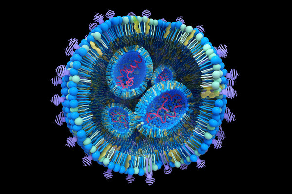
Lipid nanoparticles (LNPs) are recognised as a leading delivery platform following clinical success in vaccine and therapeutic applications. Medicines Discovery Catapult (MDC) has built a preclinical platform to evaluate LNPs against two major challenges in the field. In this blog, Lead Scientist, Dr Phil Auckland, presents a case study of this workflow using intravenously administered LNPs encapsulating mRNA, which differentially target four major liver cell types.


Blogs
15 June 2026
Preclinical evaluation of RNA nanotherapeutics – a case study of lipid nanoparticles

Opening new avenues for RNA therapeutics through a preclinical screening and optimisation platform. In this blog, Lead Scientist Dr Phil Auckland describes how applying this platform to a library of 4,500 lipid nanoparticle (LNP) formulations enabled the identification of novel candidates with desirable mechanistic properties and in vivo functionality. Furthermore, we show that integrating multiple in vitro readouts can enhance prediction and thereby improve translation.


Medicines Discovery Catapult