Opening new avenues for RNA therapeutics with a lipid nanoparticle preclinical screening and optimisation platform.
In this blog, Lead Scientist Dr Phil Auckland describes how applying this platform to a library of 4,500 lipid nanoparticle (LNP) formulations enabled the identification of novel candidates with desirable mechanistic properties and in vivo functionality. Furthermore, we show that integrating multiple in vitro readouts can enhance prediction and thereby improve translation.
June 2026
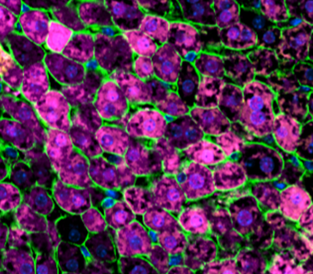
RNA therapeutics represent one of the most promising new drug modalities for addressing the challenges of modern medicine. By enabling intervention against previously intractable or “undruggable” targets, and by offering the potential for improved efficacy and safety profiles, RNA-based approaches open new avenues to treat areas of significant unmet need. Nevertheless, realising this opportunity requires advances in both the delivery technologies and the translational capabilities to assess them.
Investors and Pharma New Opportunities groups assessing RNA therapeutics and delivery platforms look to de-risk innovations across the preclinical pathway. At early stages, they prioritise clear in vitro evidence of delivery performance, strong differentiation from existing platforms and clinical benchmarks (e.g., improved targeting or tolerability), and robust assay data that validate biological claims. As programmes advance, attention shifts to convincing in vivo biodistribution and pharmacodynamic data, acceptable safety profiles at effective doses, and formulation strategies that are scalable toward GMP manufacture. Overall, they seek platforms that demonstrate robust biological validation, clear solutions to unmet delivery challenges and scalability.
Medicines Discovery Catapult, as part of the five-partner Intracellular Drug Delivery Centre (IDDC), has established an industry-standard preclinical platform to build experimental confidence in delivery vehicles for RNA therapeutics (Figure 1). Below, we describe how application of this platform to a library of 4,500 lipid nanoparticle (LNP) formulations enabled the identification of novel candidates with desirable mechanistic properties and functionality in vivo. Furthermore, we show how integration of multiple in vitro readouts can enhance prediction and therefore improve translation.
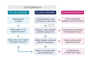
Figure 1: Assays that form the MDC preclinical cascade.
A library of 4,500 LNPs encapsulating mGreenLantern (mGL, a GFP variant) mRNA were formulated using robotic automation at the Centre for Process Innovation. This library included 125 commercial, 79 novel ionisable, and 4 novel neutral lipids, altogether spanning a large chemical and formulation space to enable the derivation of structure-activity relationships. Following physicochemical assessment of size, polydispersity, and encapsulation efficiency, the samples underwent a primary transfection screen in 384-well format benchmarked against the Moderna Spikevax SM102 formulation. Here, HeLa cells were dosed with 1ng, 10ng, and 30ng mRNA per well in triplicate, with cell confluency and mGL fluorescence intensity quantified at 48hr using high-throughput confocal microscopy. Hit formulations were identified that fulfilled target product profile criteria, of which 39 were reproduced for detailed characterisation.
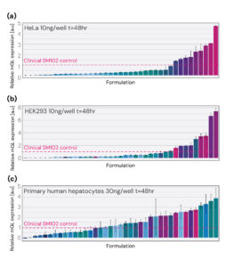
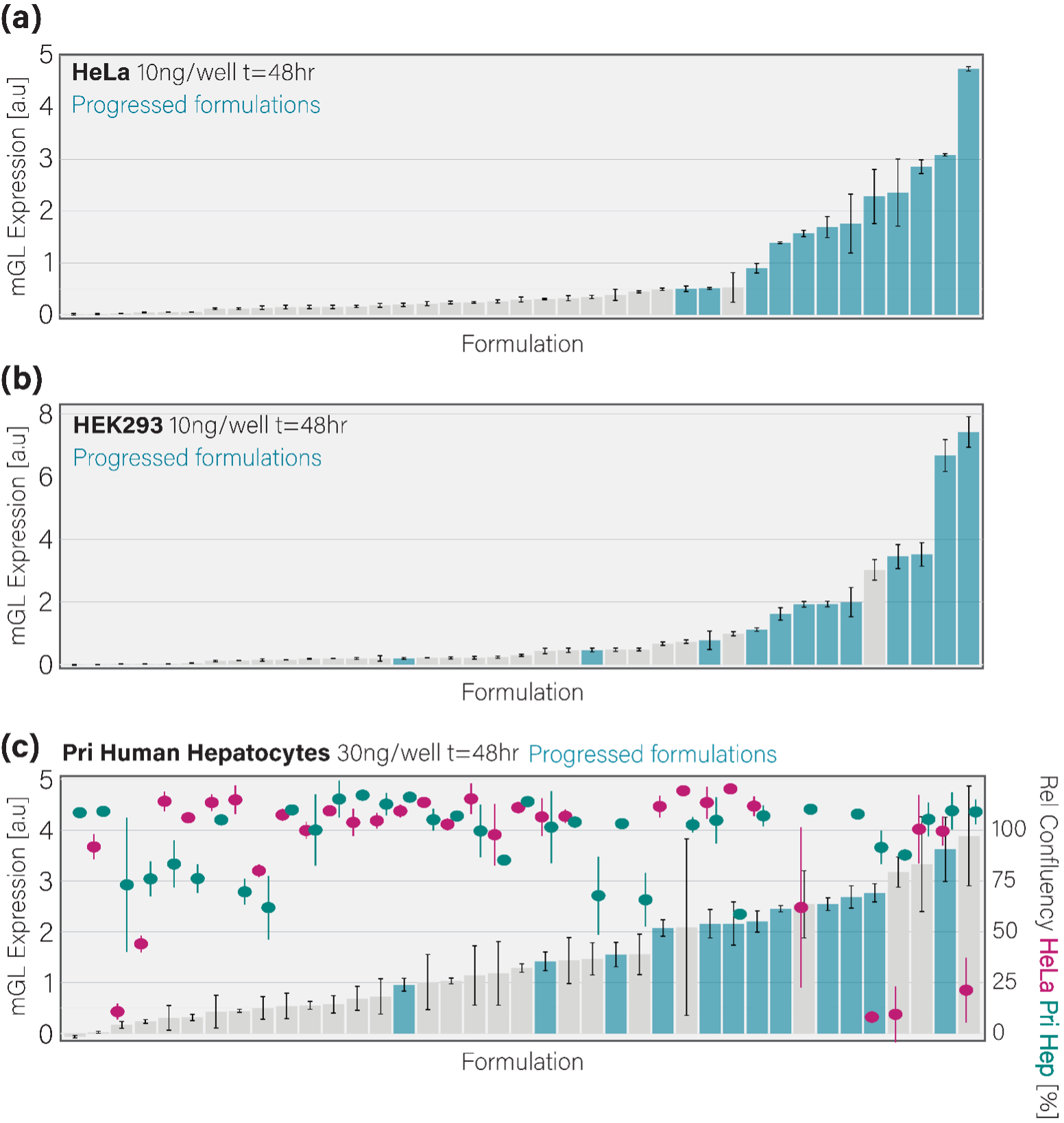
Figure 2: Assessment of potency in cell models. Relative mGL expression intensity at 48hr following dosing of HeLa cells (a), HEK293 cells (b), and primary human hepatocytes (c) with hit LNP formulations encapsulating mGL mRNA (10 or 30ng mRNA/well).
Hit formulations encapsulating mGL mRNA were dosed to HeLa cells, HEK293 cells, and primary human hepatocytes following a 7-point half-log regime. Primary hepatocytes were selected due to the propensity of intravenously administered LNPs to transfect this cell type in vivo; the cell type would be modified for formulations targeted to alternative tissues. Quantification of mGL expression at 48hr for a single dose (10 ng/well for HeLa and HEK293, 30 ng/well for primary human hepatocytes) is shown in figure 2a-c. This reveals that while high performers are conserved across cell types, an additional efficacious group of formulations are specific to primary human hepatocytes (Figure 2c).
Payload expression is an endpoint readout. For payload molecules to be expressed in target cells, LNPs must effectively internalise into endosomal vesicles, then rupture this compartment to deliver payload molecules to ribosomes in the cytosol. Functionality in vivo requires these processes to be highly efficient, as LNPs have a limited window to bind target cells, making transfection in this setting more challenging than in vitro. To identify formulations with high internalisation and rupture activity, and link this to payload expression and cellular health, we established a live-cell fluorescence-based assay. Here, payload molecules are labelled after formulation with the far-red dye SYTO60 and transfected into HeLa cells expressing Gal9-mCherry, which is specifically recruited to the intraluminal face of the limiting membrane at rupturing endosomes. This quantitative time-lapse assay allows internalisation to be visualised by SYTO60 translocation into cells, rupture to be visualised by the formation of Gal9-mCherry puncta, and payload expression to be detected by cytosolic mGL fluorescence (Figure 3a,b). Each readout had a Z-factor of ≥7.5, demonstrating a high ability to differentiate signal from background (Figure 3c). To enable comparisons across the complex dataset, maximal internalisation and rupture were combined into a normalised index metric to approximate payload delivery to the cytosol (Figure 3d). When plotted against mGL mRNA expression, this revealed formulations with the most efficient payload expression per uptake-rupture event (Figure 3d). These formulations are henceforth referred to as progressed formulations.
Progressed formulations identified through internalisation and rupture behaviour were mapped onto the 48hr cell expression data to understand predictive value (Figure 4a-c). This identified 91%, 83%, and 66% of the high-performing formulations in HeLa cells, HEK293 cells, and primary human hepatocytes, respectively (Figure 4a-c). The gap in primary hepatocyte prediction is likely due to cytotoxicity. Indeed, 50% of the primary hepatocyte-specific high-performing formulations had a significant negative effect on HeLa cell confluency while being well-tolerated by hepatocytes (Figure 4c). This demonstrates the importance of incorporating several in vitro models when optimising formulation as lipids have cell-type specific toxicity and therefore potency.
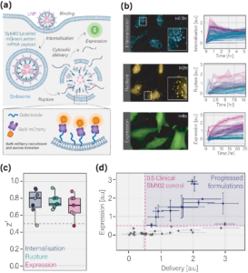
Figure 3: Understanding the dynamics of cellular internalisation, endosomal rupture and payload expression. (a) Cartoon schematic depicting the internalisation-rupture-expression assay. (b) Left: snapshots of the SYTO60, Gal9-mCherry, and mGL channels in live cells used to quantify internalisation, rupture, and expression, respectively. Right: Example time-lapse quantification of internalisation, rupture, and expression for the 39 selected formulations and controls. (c) Boxplot depicting the Z’ statistic for each repeat of the internalisation-rupture-expression assay. (d) Scatterplot of delivery (an index metric of internalisation and rupture) versus mGL expression for 39 selected formulations. Dotted pink lines indicate 0.5-fold of the SM102 clinical benchmark. Shaded area depicts the high-performing formulations progressed through the cascade.
Figure 4: Mapping delivery to expression analyses across cell models. Progressed formulations (blue bars) identified by delivery behaviour are mapped onto the 48hr mGL expression data for HeLa cells (a), HEK293 cells (b), and primary human hepatocytes (c). The 48hr confluency quantified from HeLa cells and primary human hepatocytes is additionally plotted against mGL expression for primary human hepatocytes in c.
To select formulations for testing in rodents, three criteria were set that together capture the breadth of in vitro experimental confidence: (1) >0.5 fold delivery (internalisation x rupture) in HeLa cells relative to the SM102 clinical benchmark, (2) >0.5 fold mGL expression in HeLa cells, HEK293 cells, and primary human hepatocytes relative to the SM102 clinical benchmark, and (3), a relative confluency of >85% at 48hr in HeLa cells and primary human hepatocytes (Figure 5a-e). This identified seven formulations (F1-F7), which were intravenously administered to mice via tail vein injection.
IVIS imaging of mGL expression in whole livers confirmed that all formulations except F2 were functional in vivo, with some, such as F5, outperforming the Dlin-MC3 clinical benchmark approved for siRNA delivery to the liver (Figure 6a). To validate this finding the livers were fixed, sectioned, and stained with DAPI and antibodies against mGL, CPS1 (a marker of hepatocytes) and N-Cadherin. Quantification of anti-mGL staining in single hepatocytes confirmed the IVIS observations, and suggested that F1, F6, and F7 compared positively with the Dlin-MC3 clinical benchmark (Figure 6b). The rate of in vivo functionality for the presented in vitro-in vivo cascade was 85%, compared to 53% (chance) if all 39 formulations were tested in vivo without cell-based selection.
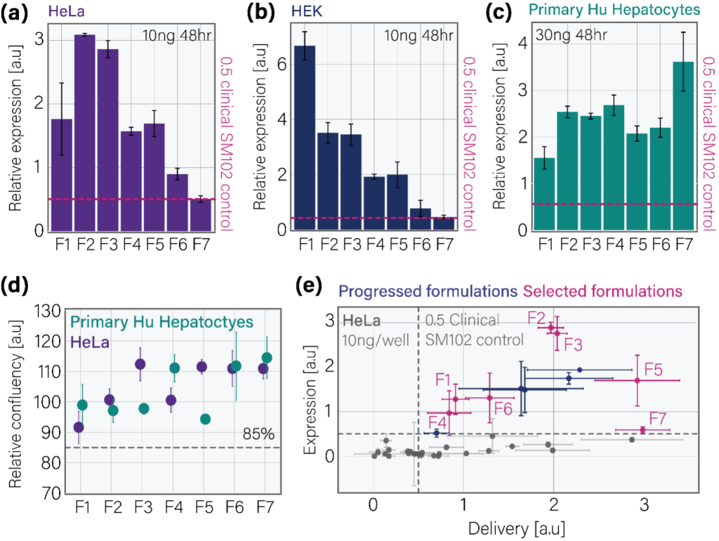
Figure 5: In vitro-based selection of candidate formulations. Formulations were selected based on the following criteria: (a-c) >0.5-fold mGL expression in HeLa cells, HEK293 cells, and primary human hepatocytes relative to clinical benchmark, (d) HeLa cell and primary human hepatocyte confluency of >85% at 48hr, and (e), a delivery-mGL expression relationship >0.5-fold that of the clinical benchmark. In all cases the benchmark is the clinical SM102 formulation.
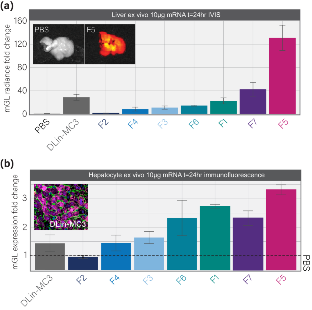
Figure 6: In vivo testing of select formulations. (a) Quantification of whole liver mGL radiance fold change using IVIS imaging. Images show the liver mGL signal for the PBS control and F5-treated mice, respectively. (b) Immunofluorescence quantification of hepatocyte mGL expression in liver sections prepared from the organs shown in (a). Image shows the staining pattern of mGL (magenta), N-cadherin (green), and CPS1 (yellow, a marker of hepatocytes) in a positive control mouse.
In summary, these data demonstrate that the Medicines Discovery Catapult platform can evaluate RNA nanotherapeutics against the central challenges facing developers. These include:
The presented workflows are not restricted to LNPs encapsulating mRNA, instead being readily applicable to other RNA nanocarriers, payload types, and advanced modalities. Through integration of this platform across preclinical programmes, MDC allows partners to generate data that informs progression against the key success criteria of right target, tissue, safety, and patient, ultimately increasing preclinical confidence and derisking the clinical path.
MDC’s preclinical cascade combines high-throughput formulation screening, mechanistic intracellular delivery characterisation, and in vivo biodistribution to build experimental confidence at every stage.
Speak to our team to find out how we can accelerate your path to the clinic.


How to study the endosomal escape of RNA therapeutics
Xenium at MDC Case Study: Hypoxia in Head and Neck Cancer
From mitochondrial networks that silently rebalance fusion and fission, to chromosomes that only reveal their fate in the minutes after division, live-cell imaging uncovers dynamics that static snapshots miss entirely. We explore three examples, including how tracking RNA therapeutic vehicles as they transit and escape endosomal compartments in real time resolves ambiguity that even super-resolution imaging of fixed cells can’t overcome.