Innovative microscopy techniques are transforming drug discovery. Technology advances are enabling the quantitative analysis of dynamic biological processes down to the nanoscale to evaluate single-molecule interactions.
High content microscopy can enable the analysis of complex multicellular structures and tissues in higher throughput than previously possible. Our range of imaging platforms enables you to explore complex biological processes, measure drug responses, and generate high-quality, decision-making data.
Visualising single cells and sub-cellular compartments supports medicines discovery by providing a non-destructive mechanistic assessment of your sample.
We have expertise in advanced fluorescence microscopy, disease specific cell biology and our scientists can draw on a diverse toolkit to characterise relationships between cell behaviour and underlying molecular processes.
Whether you are tracking drug-target engagement, investigating the mechanism of action, or validating biomarkers in cells or tissue, we offer tailored microscopy solutions that connect cellular observations to translational outcomes.
We utilise a range of advanced microscopy technologies. For therapeutic analysis, we perform different studies according to the scientific challenge:
Target validation and mechanism of action
Visualise drug-target interactions and downstream signalling using live-cell or fixed-sample assays.
Characterisation of novel therapeutics
Track binding, uptake, trafficking and release of novel drug modalities, including RNA-based therapies and novel delivery devices.
Live-cell imaging
Track biology in real-time at high resolution using live-confocal imaging.
Automated screening
Assess relevant cells in phenotypic assays at scale to support therapeutic development and screening.
Cell and tissue Biomarker analysis
Use immunofluorescence (IF), immunocytochemistry (ICC) and multiplexed imaging to analyse tissue samples, quantify expression and stratify responses.
Data analysis
Enable data interpretation using custom analysis pipelines for robust quantitative analysis of imaging data.
We have an established fluorescence microscopy platform capable of answering diverse experimental questions. This is supported by a dedicated group of imaging experts and a bioinformatics image analysis team.
Confocal microscopy
Our Zeiss LSM880 with Airyscan enables high-speed, high-resolution 4D imaging of inter- and intra-cellular events while minimising the damaging effects of laser exposure. The system is equipped with an incubator for live-cell work, assorted light sources for multiplexed imaging and an Airyscan fast mode for high-temporal resolution acquisition.
The multiphoton upgrade allows for thick samples, for example, up to 0.5 mm tissue sections, to be imaged via a specialist excitation mode.
Super-resolution microscopy
At the heart of our platform is super-resolution microscopy, powered by the Zeiss Elyra 7 system. Using single-molecule localisation microscopy (SMLM), we can image structures as small as 20 nm, well beyond the limits of conventional light microscopy.
This ultra-high-resolution approach is particularly valuable for target engagement studies where we can visualise protein-protein interactions, mechanism of action investigation and single-molecule detection in fixed cells or tissues, as well as multiplexed imaging of complex biological features.
High-content analysis
Our Opera Phenix Plus platform allows rapid, automated imaging for medium to high-throughput phenotypic analysis. With integrated liquid handling and live-cell incubation, it is ideal for therapeutic screening, functional response tracking and assay response evaluation at scale.
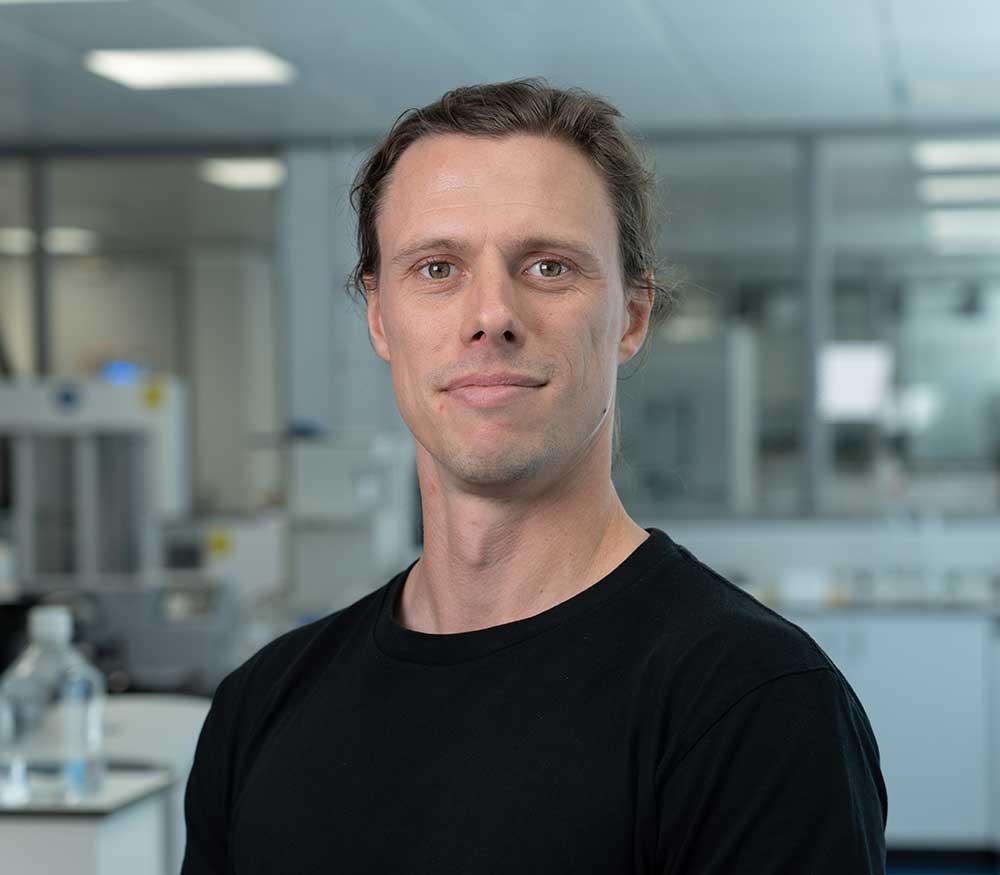
“We leverage our expertise and imaging platforms to image cells on their terms, allowing us to provide partners with straightforward conclusions that inform their medicines discovery journey. Come and speak to us and see how we can help.”
Dr Phil Auckland, Lead Scientist

Collaborate with our experts to enhance your drug discovery project. We will help you to design the right microscopy approach to answer your most important questions.
Fill in the form today and speak to us to see how we can help advance your drug discovery project.